Gaps #95115
openImage Annotation during Clinical Documentation
0%
Description
Introductory Note:
1.The Image Annotation feature in CliniCare is currently non functional . In fact, it was not adequately developed by LT to meet the requirements of clinical image annotation.
2.The “Draw from Scratch” function remains rudimentary and requires significant enhancement to support effective use.
What is body part annotation in case documentation?
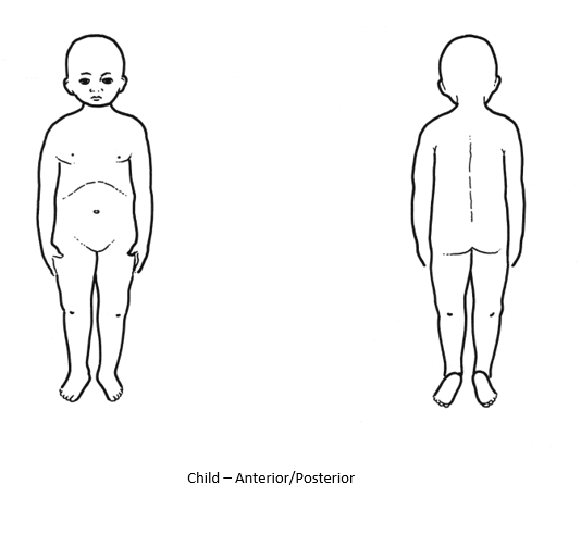
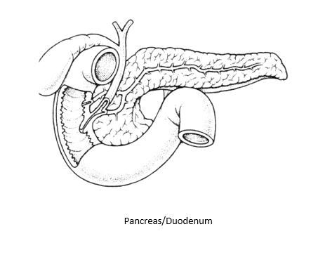
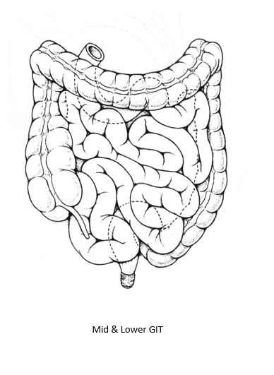
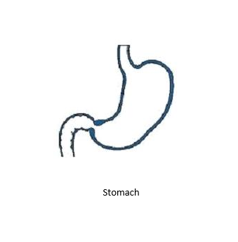
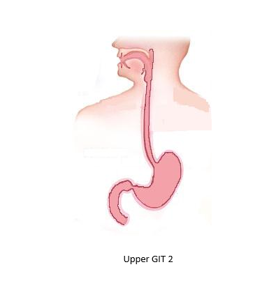
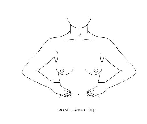
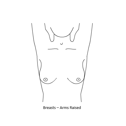
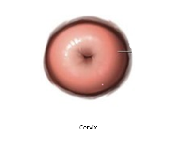
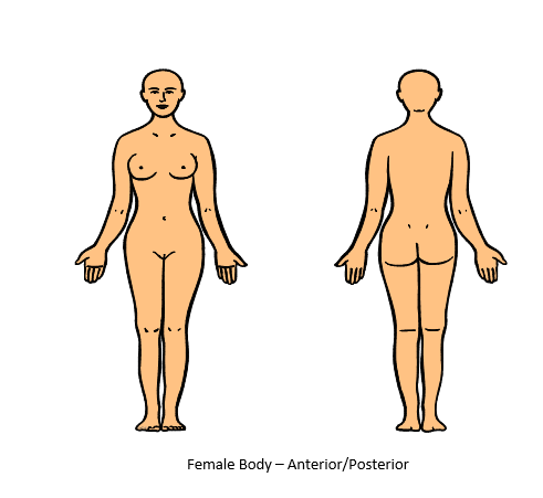
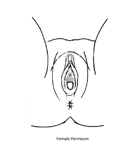
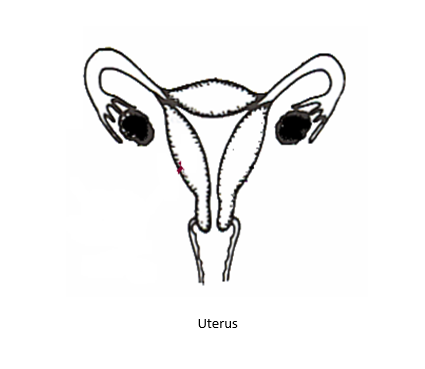
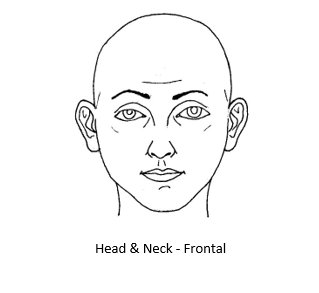
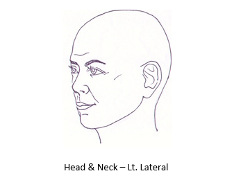
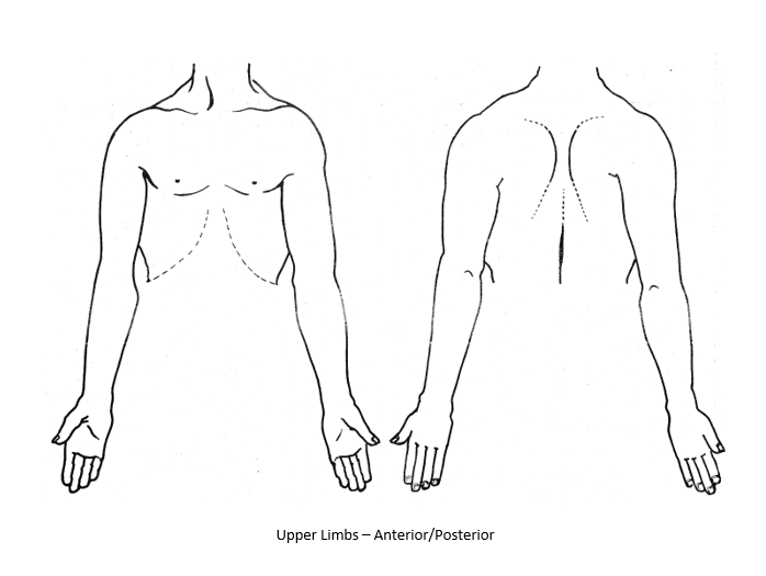
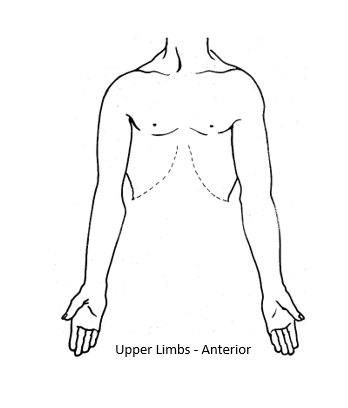
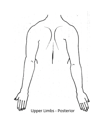
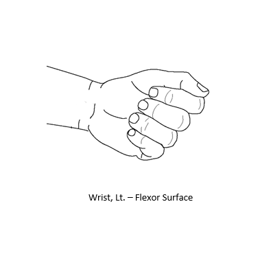
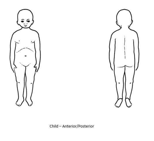
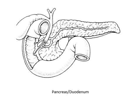
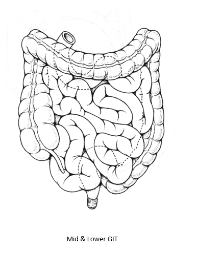
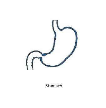
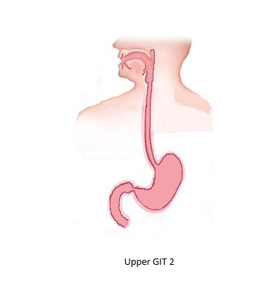
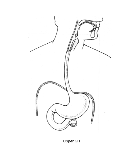
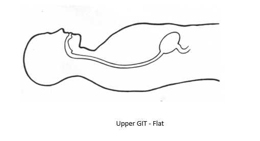
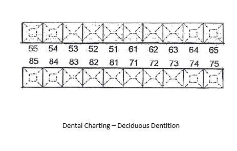
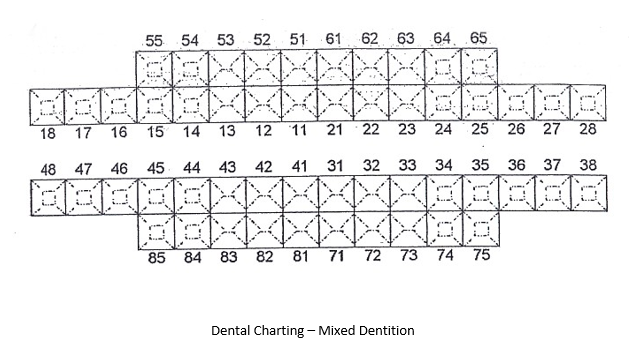
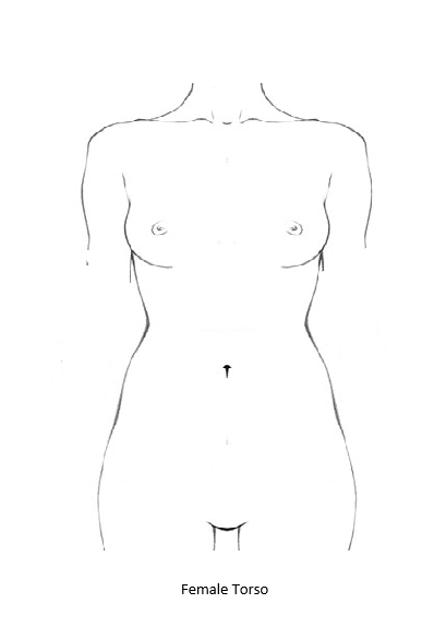
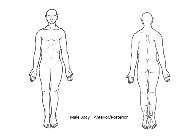
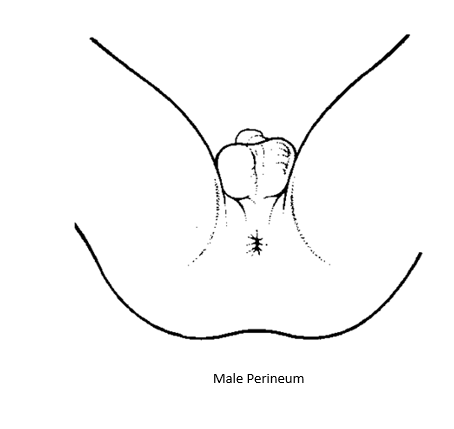
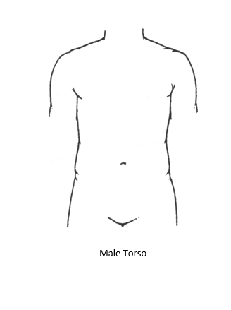
Body part annotation is a structured method for documenting injuries in trauma cases at the Emergency Department. It represents a clinical-forensic approach to injury mapping. As the adage goes, “A picture paints a thousand words” - nothing is more relevant or specific than the precise localization of injuries. Annotation shows exact injury sites, reducing ambiguity in narrative notes.
In trauma documentation, body part annotation transforms injury records into structured, standardized, and legally defensible evidence. It ensures that every wound is precisely located and accurately described, supporting both clinical care and medico legal accountability.
Medico Legal Perspective
• Trauma documentation is often court admissible evidence.
• In cases of assault, abuse, or accidents, precise injury mapping supports forensic analysis.
• Consistency between ED notes, radiology reports, and surgical findings is critical to prevent discrepancies, ensure accuracy, maintain clinical integrity, and uphold medico legal accountability in trauma case documentation
Use Case: Application of Body Part Annotation in Trauma Documentation
1. A patient presents with a 3 cm lacerated wound at the center of the right palm.
2. The clinician retrieves the image of the right palm from the system.
3. The clinician marks the injury at its exact location, enters the wound type, and defines the wound length.
4. The annotated image is saved into the system.
5. The annotated image is stored in two locations:
(a) within the patient’s case notes
(b) in the designated body part placeholder.
Files
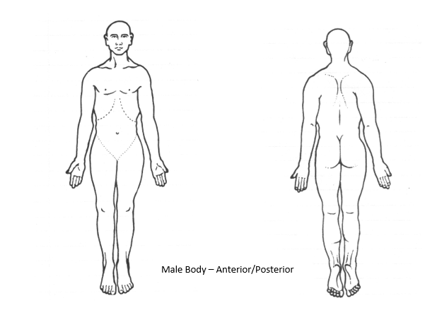
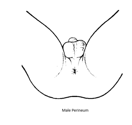
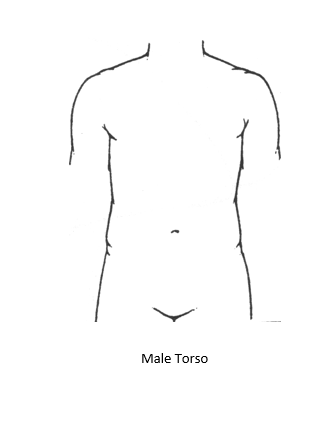
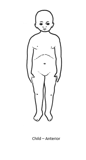
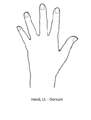
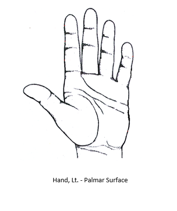
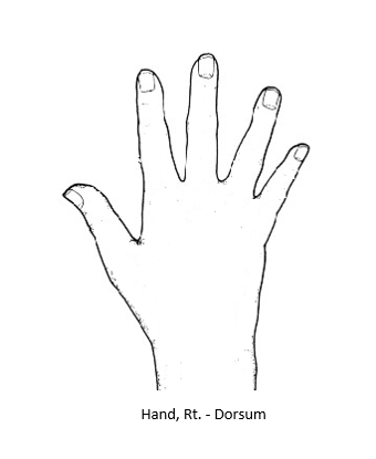
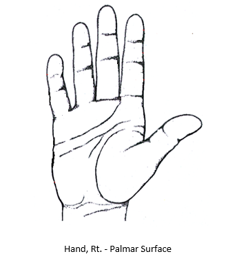
Updated by CP Chan 3 months ago
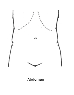
- File Abdomen.PNG Abdomen.PNG added
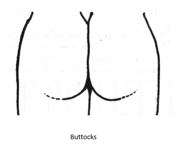
- File Buttocks.PNG Buttocks.PNG added
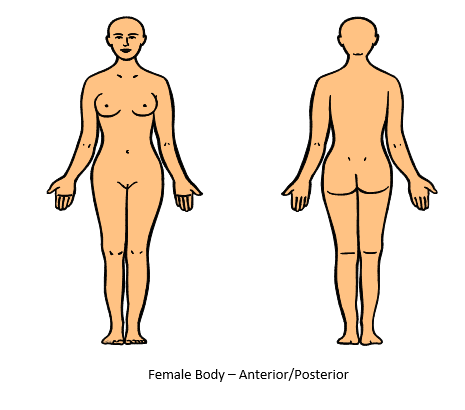
- File Female Body-Anterior and Posterior.PNG Female Body-Anterior and Posterior.PNG added
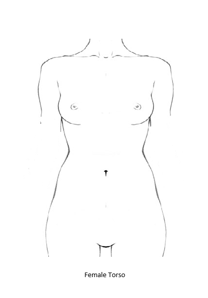
- File Female Torso.PNG Female Torso.PNG added
- File Male Body-Anterior and Posterior.PNG Male Body-Anterior and Posterior.PNG added
- File Male Perineum.PNG Male Perineum.PNG added
- File Male Torso.PNG Male Torso.PNG added
- File Child-Anterior and Posterior.PNG Child-Anterior and Posterior.PNG added
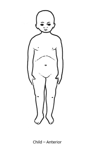
- File Child-Anterior.PNG Child-Anterior.PNG added
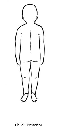
- File Child-Posterior.PNG Child-Posterior.PNG added
Updated by CP Chan 3 months ago
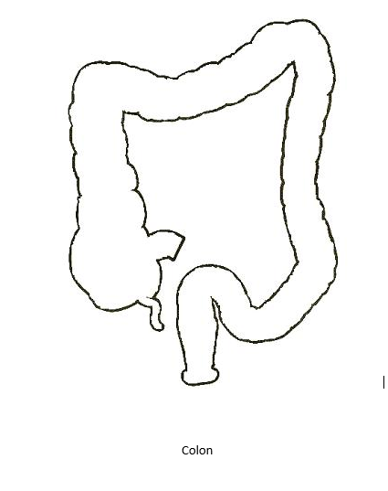
- File Colon.PNG Colon.PNG added
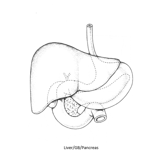
- File Liver-GB-Pancreas.PNG Liver-GB-Pancreas.PNG added
- File Mid and Lower GIT.PNG Mid and Lower GIT.PNG added
- File Pancreas-Duodenum.PNG Pancreas-Duodenum.PNG added
- File Stomach.PNG Stomach.PNG added
- File Upper GIT 2.PNG Upper GIT 2.PNG added
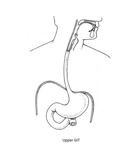
- File Upper GIT.PNG Upper GIT.PNG added
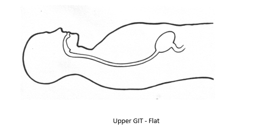
- File Upper GIT-Flat.PNG Upper GIT-Flat.PNG added
Updated by CP Chan 3 months ago
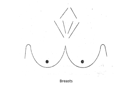
- File Breasts.PNG Breasts.PNG added
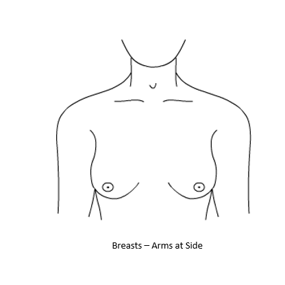
- File Breasts-Arms at Side.PNG Breasts-Arms at Side.PNG added
- File Breasts-Arms on Hips.PNG Breasts-Arms on Hips.PNG added
- File Breasts-Arms Raised.PNG Breasts-Arms Raised.PNG added
- File Cervix.PNG Cervix.PNG added
- File Female Body-Anterior and Posterior.PNG Female Body-Anterior and Posterior.PNG added
- File Female Perineum.PNG Female Perineum.PNG added
- File Uterus.PNG Uterus.PNG added
Gynae Images
Updated by CP Chan 3 months ago
- File Head and Neck-Frontal.PNG Head and Neck-Frontal.PNG added
- File Head and Neck-Lt Lateral.PNG Head and Neck-Lt Lateral.PNG added
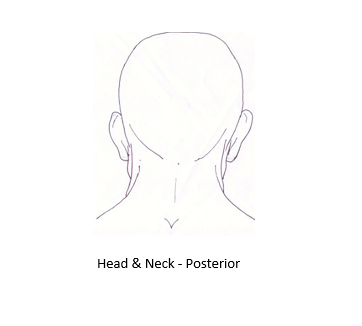
- File Head and Neck-Posterior.PNG Head and Neck-Posterior.PNG added
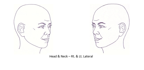
- File Head and Neck-Rt and Lt Lateral.PNG Head and Neck-Rt and Lt Lateral.PNG added
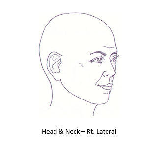
- File Head and Neck-Rt Lateral.PNG Head and Neck-Rt Lateral.PNG added
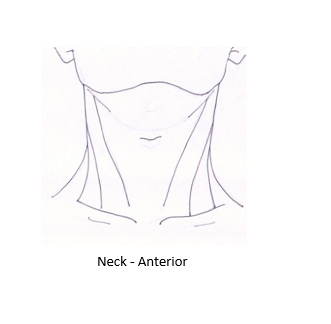
- File Neck-Anterior.PNG Neck-Anterior.PNG added
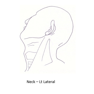
- File Neck-Lt Lateral.PNG Neck-Lt Lateral.PNG added
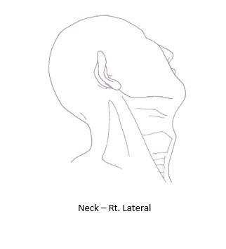
- File Neck-Rt Lateral.PNG Neck-Rt Lateral.PNG added
Head & Neck Images
Updated by CP Chan 3 months ago
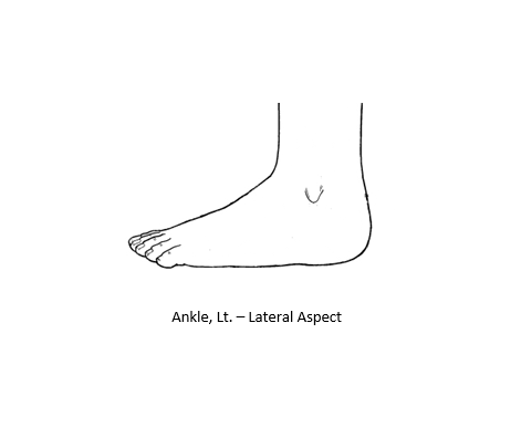
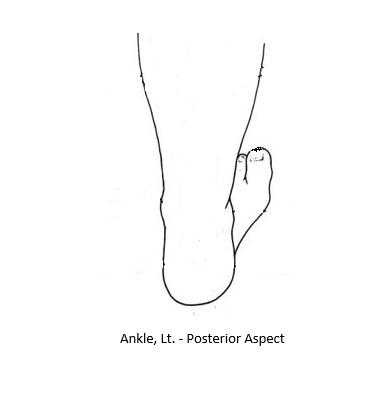
- File Ankle Lt-Lateral Aspect.PNG Ankle Lt-Lateral Aspect.PNG added
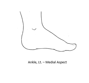
- File Ankle Lt-Medial Aspect.PNG Ankle Lt-Medial Aspect.PNG added
- File Ankle Lt-Posterior Aspect.PNG Ankle Lt-Posterior Aspect.PNG added
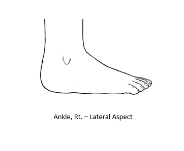
- File Ankle Rt-Lateral Aspect.PNG Ankle Rt-Lateral Aspect.PNG added
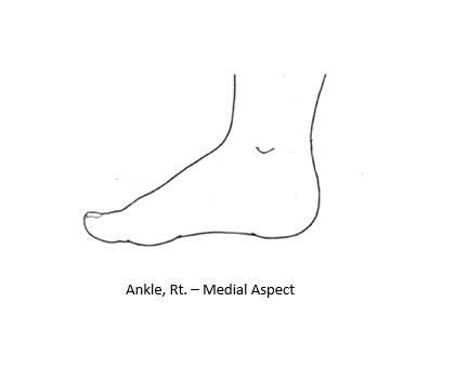
- File Ankle Rt-Medial Aspect.PNG Ankle Rt-Medial Aspect.PNG added
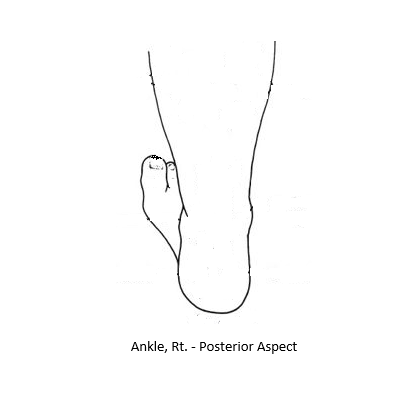
- File Ankle Rt-Posterior Aspect.PNG Ankle Rt-Posterior Aspect.PNG added
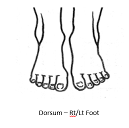
- File Dorsum-Rt and Lt Foot.PNG Dorsum-Rt and Lt Foot.PNG added
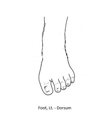
- File Foot Lt-Dorsum.PNG Foot Lt-Dorsum.PNG added
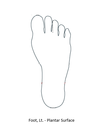
- File Foot Lt-Plantar Surface.PNG Foot Lt-Plantar Surface.PNG added
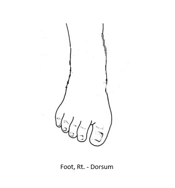
- File Foot Rt-Dorsum.PNG Foot Rt-Dorsum.PNG added
LOwer Limb Images
Updated by CP Chan 3 months ago
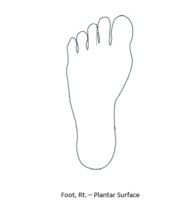
- File Foot Rt-Plantar Surface.PNG Foot Rt-Plantar Surface.PNG added
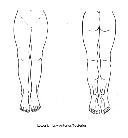
- File Lower Limbs-Anterior and Posterior.PNG Lower Limbs-Anterior and Posterior.PNG added
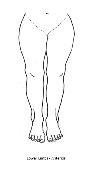
- File Lower Limbs-Anterior.PNG Lower Limbs-Anterior.PNG added
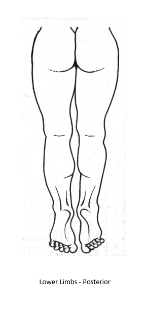
- File Lower Limbs-Posterior.PNG Lower Limbs-Posterior.PNG added
Lower Limb Images, Contd
Updated by CP Chan 3 months ago
- File Hand Lt-Dorsum.PNG Hand Lt-Dorsum.PNG added
- File Hand Lt-Palmer Surface.PNG Hand Lt-Palmer Surface.PNG added
- File Hand Rt-Dorsum.PNG Hand Rt-Dorsum.PNG added
- File Hand Rt-Palmar Surface.PNG Hand Rt-Palmar Surface.PNG added
- File Upper Limbs-Anterior and Posterior.PNG Upper Limbs-Anterior and Posterior.PNG added
- File Upper Limbs-Anterior.PNG Upper Limbs-Anterior.PNG added
- File Upper Limbs-Posterior.PNG Upper Limbs-Posterior.PNG added
- File Wrist Lt-Flexor Surface.PNG Wrist Lt-Flexor Surface.PNG added
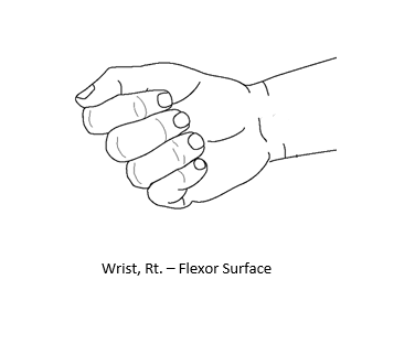
- File Wrist Rt-Flexor Surface.PNG Wrist Rt-Flexor Surface.PNG added
Upper Limb Images
Updated by CP Chan 3 months ago
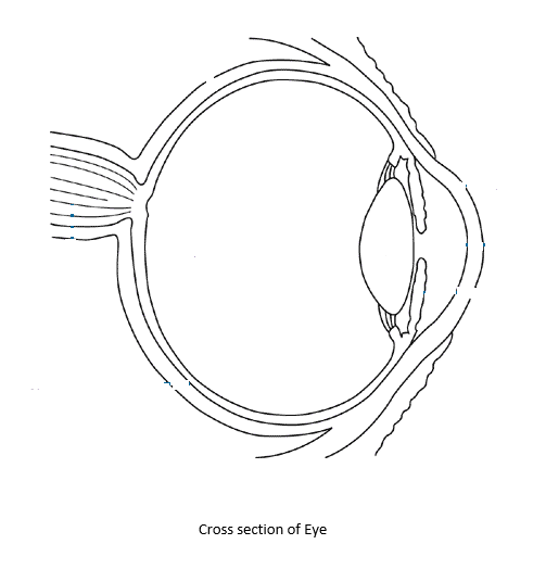
- File Cross section of Eye.PNG Cross section of Eye.PNG added
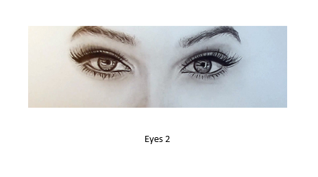
- File Eyes 2.PNG Eyes 2.PNG added
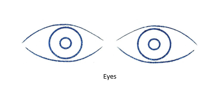
- File Eyes.PNG Eyes.PNG added
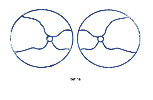
- File Retina.PNG Retina.PNG added
Ophthalmology Images
Updated by CP Chan 3 months ago
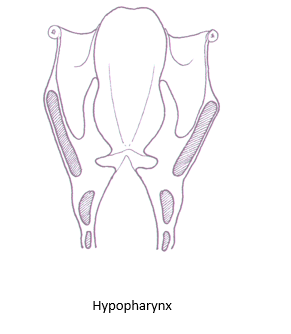
- File Hypopharynx.PNG Hypopharynx.PNG added
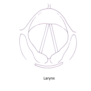
- File Larynx.PNG Larynx.PNG added
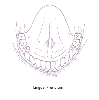
- File Lingual Frenulum.PNG Lingual Frenulum.PNG added
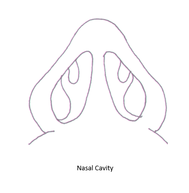
- File Nasal Cavity.PNG Nasal Cavity.PNG added
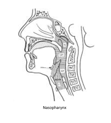
- File Nasopharynx.PNG Nasopharynx.PNG added
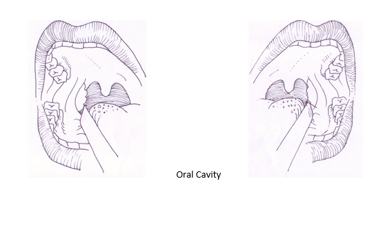
- File Oral Cavity.PNG Oral Cavity.PNG added
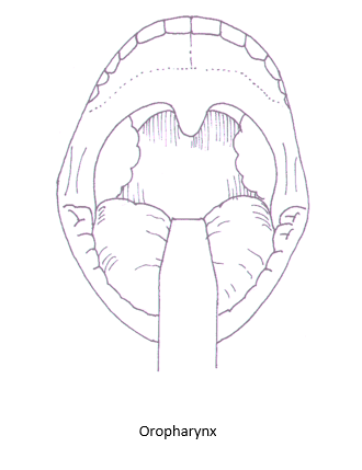
- File Oropharynx.PNG Oropharynx.PNG added
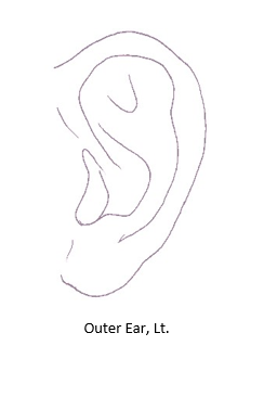
- File Outer Ear, Lt.PNG Outer Ear, Lt.PNG added
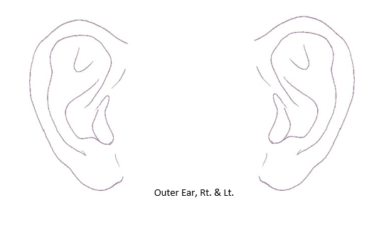
- File Outer Ear, Rt and Lt.PNG Outer Ear, Rt and Lt.PNG added
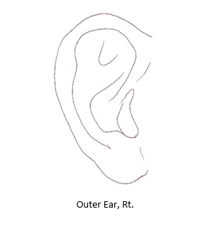
- File Outer Ear, Rt.PNG Outer Ear, Rt.PNG added
Otorhinolaryngology Images
Updated by CP Chan 3 months ago
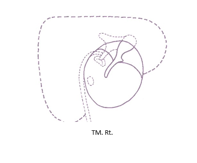
- File TM Rt.PNG TM Rt.PNG added
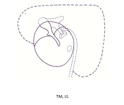
- File TM, Lt.PNG TM, Lt.PNG added
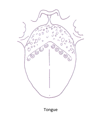
- File Tongue.PNG Tongue.PNG added
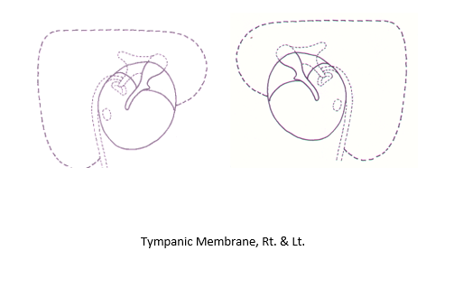
- File Tympanic Membrane, Rt and Lt.PNG Tympanic Membrane, Rt and Lt.PNG added
Otorhinolaryngology Images, Continue
Updated by CP Chan 3 months ago
- File Annotation Screens.docx Annotation Screens.docx added
Annotation Screens
Updated by CP Chan 3 months ago
- File Abdomen.PNG Abdomen.PNG added
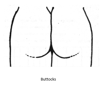
- File Buttocks.PNG Buttocks.PNG added
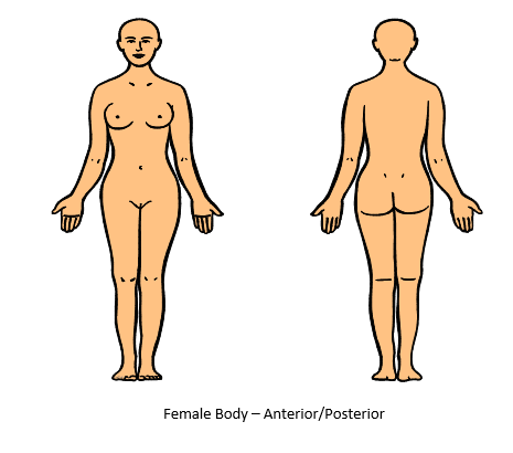
- File Female Body-Anterior and Posterior.PNG Female Body-Anterior and Posterior.PNG added
- File Female Torso.PNG Female Torso.PNG added
- File Male Body-Anterior and Posterior.PNG Male Body-Anterior and Posterior.PNG added
- File Male Perineum.PNG Male Perineum.PNG added
- File Male Torso.PNG Male Torso.PNG added
Adult Body Images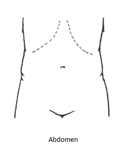
Updated by CP Chan 3 months ago
- File Child-Anterior and Posterior.PNG Child-Anterior and Posterior.PNG added
- File Child-Anterior.PNG Child-Anterior.PNG added
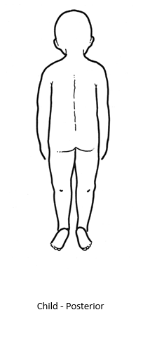
- File Child-Posterior.PNG Child-Posterior.PNG added
Child Body Images
Updated by CP Chan 3 months ago
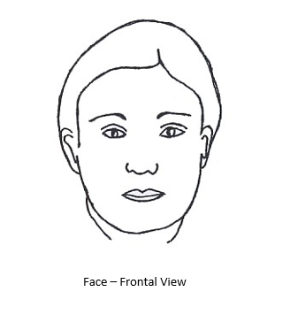
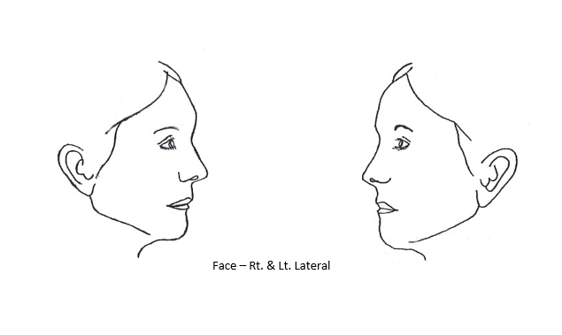
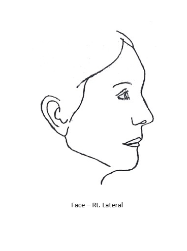
- File Face-Frontal View.PNG Face-Frontal View.PNG added
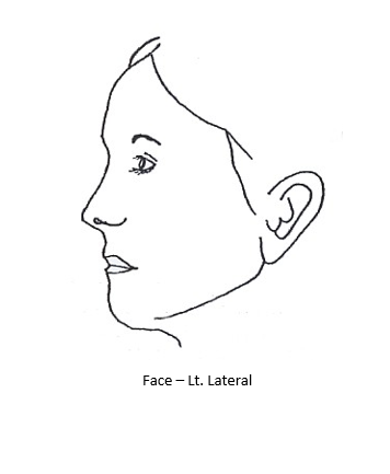
- File Face-Lt Lateral.PNG Face-Lt Lateral.PNG added
- File Face-Rt and Lt Lateral.PNG Face-Rt and Lt Lateral.PNG added
- File Face-Rt Lateral.PNG Face-Rt Lateral.PNG added
Face Images
Updated by CP Chan 3 months ago
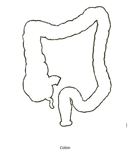
- File Colon.PNG Colon.PNG added
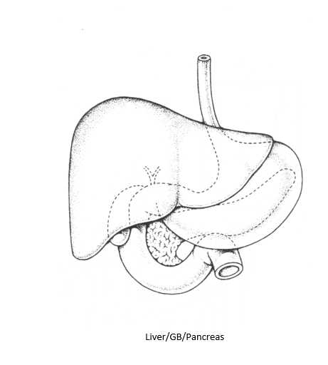
- File Liver-GB-Pancreas.PNG Liver-GB-Pancreas.PNG added
- File Mid and Lower GIT.PNG Mid and Lower GIT.PNG added
- File Pancreas-Duodenum.PNG Pancreas-Duodenum.PNG added
- File Stomach.PNG Stomach.PNG added
- File Upper GIT 2.PNG Upper GIT 2.PNG added
- File Upper GIT.PNG Upper GIT.PNG added
- File Upper GIT-Flat.PNG Upper GIT-Flat.PNG added
GIT Images
Updated by CP Chan 3 months ago
- File Dental Charting-Deciduous Dentition.PNG Dental Charting-Deciduous Dentition.PNG added
- File Dental Charting-Mixed Dentition.PNG Dental Charting-Mixed Dentition.PNG added
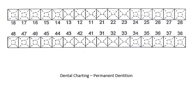
- File Dental Charting-Permanent Dentition.PNG Dental Charting-Permanent Dentition.PNG added
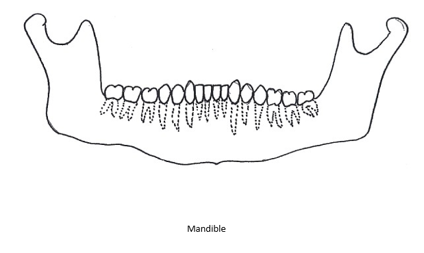
- File Mandible.PNG Mandible.PNG added
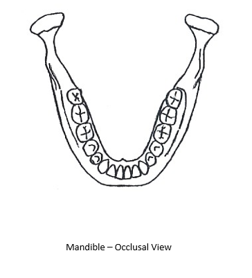
- File Mandible-Occlusal View.PNG Mandible-Occlusal View.PNG added
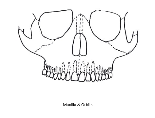
- File Maxilla and Orbits.PNG Maxilla and Orbits.PNG added
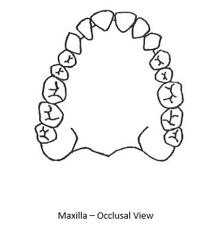
- File Maxilla-Occlusal View.PNG Maxilla-Occlusal View.PNG added
Dental Images